

Retinal detachment

We operate on retinal detachment to prevent blindness in the affected eye. Our team of specialists puts their knowledge and experience at the patient’s service to provide the quality care you need with advanced techniques and technologies.

Older man covering his eye

What is retinal detachment?

In retinal detachment, a separation occurs between the neurosensory retina and the pigment epithelium when a tear forms through which fluid passes from the vitreous cavity into the subretinal space.

What is the treatment?

Retinal detachment is treated with surgery. An extensive vitrectomy is performed, the retina is reattached using heavy liquids, and then laser treatment is applied around the breaks, leaving the eyeball filled with gas or silicone oil in complex cases.

Vitrectomy can be combined with the placement of a scleral buckle, which allows us to increase the surgical success rate even further.

At Instituto Castanera, after a thorough study of your case, the specialist will prescribe the appropriate treatment. We have advanced equipment and a team with years of experience that is constantly updating its knowledge to apply the most innovative and effective techniques. We provide you with the professional and caring attention that helps you feel at ease throughout the entire process.

Our specialists talk about retinal detachment

Retina and Vitreous – Floaters
Dr. Laura Broc, a retina and vitreous specialist at Instituto de Oftalmología Castanera, explains what eye floaters (myodesopsias) are and which symptoms should alert the patient.

Professionals who form the medical team for retinal detachment

Medical license no.: 33258 COMB

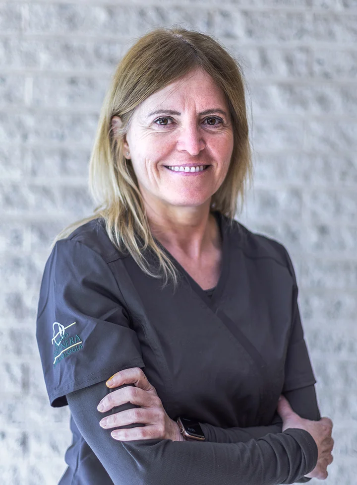
Dr. Laura Broc Iturralde

Medical license no.: 33258 COMB

Dr. Laura Broc Iturralde

Graduated in Medicine and Surgery from the University of Barcelona in 1996 and specialist in Ophthalmology since 2002. Master’s degree in Vitreoretinal Disease from the UAB, where she is an associate professor.

Her work at Instituto Castanera focuses on the medical and surgical diagnosis and treatment of retinal diseases. She takes part in clinical trials investigating the efficacy of different drugs for the prevention and treatment of age-related macular degeneration.

Medical Societies:

  • Catalan Society of Ophthalmology
  • Spanish Society of Ophthalmology (S.E.O)
  • Spanish Society of Retina and Vitreous (S.E.R.V), board member

Frequently asked questions

Retinal detachments cause a loss of visual field that increases over the course of several days. They are usually preceded by myodesopsia (“floaters”) and photopsia (“flashes”) during the days before the detachment occurs.

Most likely not, as long as the detachment is recent. Fortunately, in recent years vitreoretinal surgery has advanced enormously, achieving successful results in most cases.

In general, it is not painful. If a scleral buckle is also placed, the eye may be somewhat more inflamed during the first few days after surgery. One month after the procedure, the eye looks completely normal.

No. Generally, the patient must remain face down during the first week after surgery. If a short-acting gas is used, it takes around 15 days to be reabsorbed, and during this period vision is not sharp. The patient must take relative rest during the first month after surgery. Visual recovery is gradual over the course of several months.

Phone:

+34 932 173 704

Language: